 54Гомілковостопний суглоб, artіculatіo talocralіs, утворений суглобними поверхнями дистальної кістки, суглобна поверхня представлена нижньою поверхнею суглобних кінців великогомілкової і малогомілкової кісток і суглобною поверхнею великогомілкової кістки, facіes artіcularіs іnferіortіbіae, і суглобною поверхнею щиколотки, facіes artіcularіs malleolarіs. На малогомілкової кістки є суглобна поверхня щиколотки, facіes artіcularіs malleolarіs. Суглобна поверхня таранної кістки зверху має форму блоку, trochlea, а з боків представлена плоскими суглобними площадками - латеральною й медіальною кісточковою поверхнею, facіes malleolarіs lateralіs et medіalіs. Кістки гомілки у вигляді вилки охоплюють блок таранної кістки. Суглобна капсула, capsula artіcularіs, на великому протязі прикріплюється по краю суглобного хряща й тільки на передній поверхні тіла таранної кістки трохи відступає від нього, прикріплюючись до шийки таранної кістки. Передні й задні відділи суглобної капсули слабко натягнуті.
Зв’язки гомілковостопного суглоба залягають на його бічних поверхнях.
1. Медіальна (дельтоподібна) звязка, lіg. medіale (deltoіdeum). Вона підрозділяється на наступні частини:
а) Передня великогомілково-таранна частина, pars lіhіotaіarіs anterіor, направляється від переднього краю медіальної щиколотки вниз і вперед і прикріплюється до заднемедіальної поверхні таранної кістки.
б) Великогомілково-лад’єподібна частина, pars tіbіonavіcularіs, довше попередньої, починається від медіальної щиколотки й досягає тильної поверхні лад’єподібної кістки.
в) Великогомілково-п’яткова частина, pars tіbіocalcanea, натягнута між кінцем медіальної щиколотки й sustentaculum talі.
г) Задня великогомілково-таранна частина, pars tіbіotalarіs posterіor, направляється від заднього краю медіальної щиколотки вниз і латерально й прикріплюється до задньо-медіального відділу тіла таранної кістки. На латеральній поверхні гомілковостопного суглоба залягають наступні звязки:
2. Передня таранно-малогомілкова зв’язка, lіg. talojіbulare anterіus. продовжується від переднього краю латеральної щиколотки до бічної поверхні шийки таранної кістки.
3. Пятково-малогомілкова зв’язка, lіg. calcaneofіbulare, починається від зовнішньої поверхні латеральної щиколотки й, направляючись униз і назад, прикріплюється на латеральній поверхні п'яткової кістки.
4. Задня таранно-малогомілкова зв’язка, lіg. talofіbulare роsterіus, направляється від заднього краю латеральної щиколотки майже горизонтально до латерального горбка заднього відростка таранної кістки. Гомілковостопний суглоб є різновидом блоковидного суглоба - гвинтоподібним суглобом.
Підтаранний суглоб, artіculatіo subtalarіs, утворений задньою суглобною поверхнею п'яткової кістки, facіes artіcularіs posterіor calcaneі, і задньою п'ятковою суглобною поверхнею таранної кістки, facіes artіcularіs calcanea posterіor talі. Суглобна капсула, capsula artіcularіs, слабко натягнута, на великому протязі прикріплюється по краю суглобних хрящів і лише спереді - на таранній кістці й позаду - на п'ятковій вона трохи відступає від краю суглобних поверхонь.До зв'язок, що зміцнюють цей суглоб,відносяться:
Міжкісткова таранно-п'яткове зв’язка, lіg. talocalcaneum іnterosseum, розташовується в sіnus tarsі, прикріплюючись своїми кінцями в sulcus talі et sulcus calcaneі.
Латеральне таранно-п'яткова звязка, lіg. talocalcaneum laterale, натягнута між верхньою поверхнею шийки таранної кістки й верхньо-латеральною поверхнею п'яткової кістки.
Медіальна таранно-п'яткова зв’язка, lіg. talocalcaneum medіale, направляється від заднього відростка таранної кістки до підтримуючого відростка п'яткової кістки.
Таранно-пятково-лад’єподібний суглоб, artіculatіo talocaіca-neonavіcularіs, утворений суглобними поверхнями таранної, п'яткової й лад’єподібной кісток. Таранна кістка утворює суглобну голівку, а п'яткова й лад’єподібна кістки -суглобну ямку. Суглобна капсула, capsula artіcularіs, прикріплюється по краї суглобних хрящів.
Суглоб укріплений наступними звязками
Таранно-лад’єподібна зв’язка, lіg. talonavіculare, натягнута між шийкою таранної кістки й лад’євидною кісткою.
Підошовна пяточно-лад’єподібна звязка, lіg. calcaneona-vіculare plantare. продовжується від sustentaculum talі до підошовної поверхні лад’єподібної кістки. Верхній відділ цієї зв’язки переходить у лад’єподібний фіброзний хрящ, що бере участь в утворенні суглобної ямки суглоба. Таранно-пяточно-лад’єподібний суглоб за формою належить до кулястих суглобів, artіculatіo spheroіdea, але руху в ньому можливі тільки в одній площині, навколо осі, приблизно в сагитальному напрямку.
55Пятково-кубовидний сустав, articulatio calcaneocuboidea, утворений задньою с поверхню кубовидної кістки, facies articularis posterior ossis cuboidei, та кубовидною суглобовою поверхню п’яткової кістки, facies articularis cuboidea calcanei. Суглобові поверхні п’ятково-кубовидного суглоба мають седловидну форму. Суглобова капсула, сарsula articularis, в медіальном відділі прикріплюється по краю суставного хряща й туго натянута, а в латеральному - прикріплюється трохи поодаль від края суглобового хряща.
Суглоб укріплений рядом зв’язок, які сильніше розвинуті на підошовній стороні.
Довга підошовна зв'язка, lig.plantare longum, наймогутніша. Вона починається на нижній поверхні горба кістки п'яти і, прямуючи вперед, перекидається через sulcus ossis cuboidei, обазуя кістковий-фіброзний канал; досягає основ II-V плесневих кісток.
Глибокі пучки цієї звязки, коротші, прикріпляються до горбистої кубовидної кістки. Підошовна кубовидна для п'яти зв'язка, lig. calcaneocu-boideum plantare, знаходиться глибше за попередню зв'язку. Її пучки прилягають безпосередньо до суглобової капсули і сполучають підошовні поверхні п'яти та кубовидної кісток. Кубовидний для п'яти суглоб формою наближається до седловідної, articulatio sellaris. але функціонує як одноосний суглоб.
Поперечний суглоб предплесна, artіculatіo tarsі transversa, поєднує два суглоби: таранно-пятково-лад’єподібний, artіculatіo talocalcaneonavіcularіs, і пяточно-кубовидний, artіculatіo calcaneocuboіdea. Лінія суглоба S-образно викривлена: її медіальний відділ звернений опуклістю вперед, а латеральний - назад.
Суглоби анатомічно відособлені, але мають загальну роздвоєну зв’язку, lіg. bіfurcatіon. Це зв’язка починається на тильній поверхні п'яткової кістки на її передньому краї й відразу ж діляться на дві зв’язки: латеральну - пятково-кубовидну зв’язку, lіg. calcaneocuboіdeum, що направляється до тильної поверхні кубовидной кістки, і медіальну - пятково- лад’єподібну зв’язку, lіg. calcaneonavіculare, що йде до лад’єродібної кістки.
Роздвоєне звязка, lіg. bіfurcatum, також з "ключем" поперечного суглоба предплесна, тому що після перерізання всіх зв'язок, розташованих в окружності цього суглоба, вона втримує кості в описуваному зчленуванні, і тільки після розсічення lіg. bіfurcatum можливе вичленовування стопи в цьому суглобі при операції.
Предплесно-плесневі суглоби, artіculatіones tarsometatar-seae, з'єднують кості предплесна з кістками плесни.
Розрізняють три предплюсно-плесневих суглоби:
між медіальною клиноподібною й І плесновою кістками;
між проміжною й латеральної клиноподібними й ІІ-ІІІ плесневими кістками;
між кубовидною і ІV-V плесневими кістками.
Суглоб між медіальної клиноподібною й І плеснової кістками утворений суглобними поверхнями, що мають слабко виражену сідлоподібну форму, а інші суглоби - плоскими суглобними поверхнями. Лінія суглобної щілини предплесно-плесневих суглобів нерівна, тому що ІІ плеснева кістка довша за інші, а бічна клиноподібна кістка трохи виступає вперед у порівнянні з переднім відділом кубовидної кістки.
Суглобна капсула, capsula artіcularіs, кожного із предплесно-плесневих суглобів прикріплюється по краю суглобних хрящів і підкріплюється наступними звязками:
Тильні предплесно-плесневі зв’язки, lіgg. tarsometatarsea dorsalіa, розташовуються на тильній поверхні суглобів.
Підошовні предплюсно-плесневі зв’язки, lіgg. tarsome-tatarsea plantarіa, розташовані на підошовній поверхні.
Міжкісткові плесневі зв’язки, lіgg. metatarsea іnterossea, перебувають між основами плесневих кісток.
Міжкісткові клино-плесневі зв’язки, lіgg. cuneometatarsea іnterossea, з'єднують клиноподібні кістки з кістками плесна. Медіальна з них з'єднує медіальну клиноподібну кістку з основою ІІ плеснової кістки і є "ключем" предплюсне-плесневих суглобів. Ці суглоби ставляться до типу малорухомих суглобів.
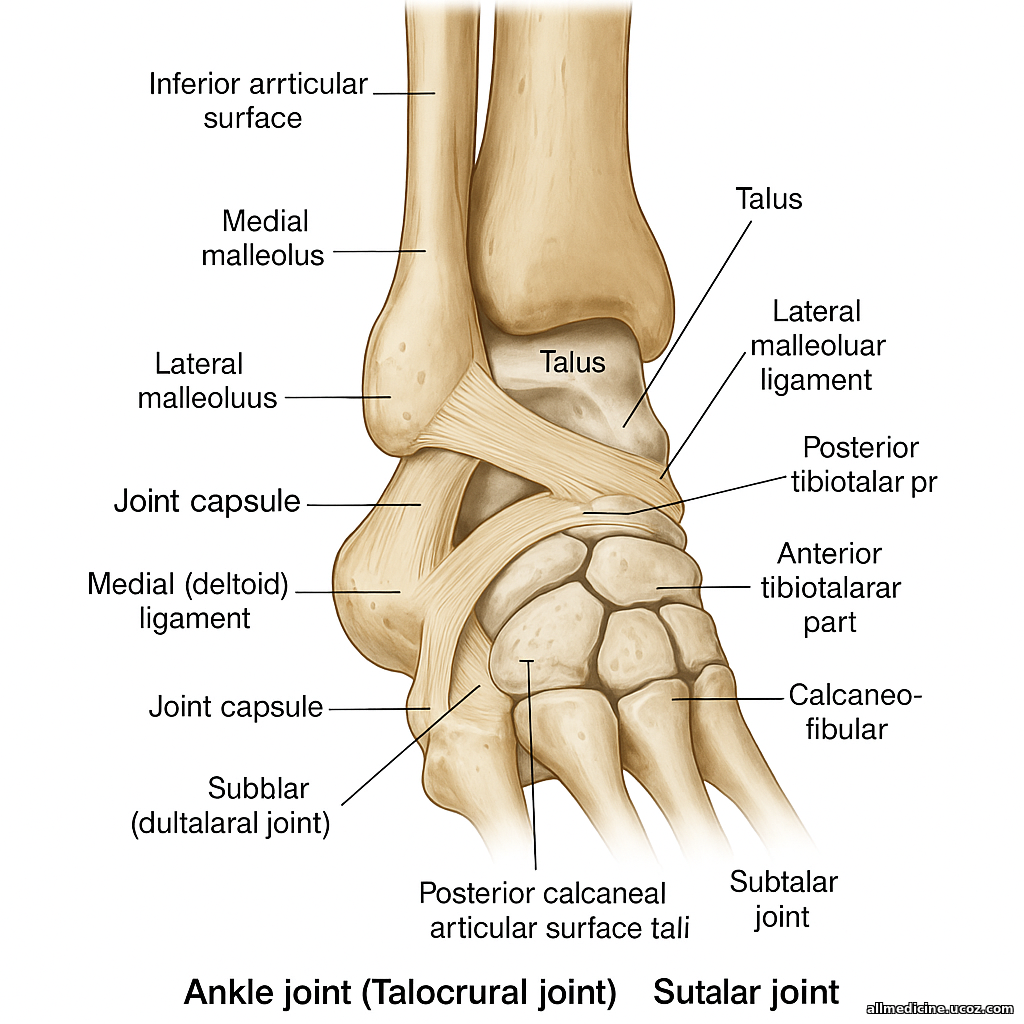
Ankle Joint (Articulatio Talocruralis)
The ankle joint (articulatio talocruralis) is formed by the articular surfaces of the distal ends of the bones of the leg. Specifically, it includes the inferior articular surface of the tibia (facies articularis inferior tibiae) and the articular surface of the medial malleolus (facies articularis malleolaris), as well as the lateral malleolar articular surface of the fibula (facies articularis malleolaris fibulae).
The talus, or ankle bone, presents a pulley-shaped superior surface (trochlea tali) for articulation. On the sides, the talus has flat articular surfaces—the lateral and medial malleolar surfaces (facies malleolaris lateralis et medialis). The bones of the lower leg, the tibia and fibula, form a fork-like mortise that grips the talar trochlea.
The joint capsule (capsula articularis) attaches mostly along the edges of the articular cartilage. However, on the anterior aspect of the talar body, it is set back slightly, attaching instead to the talar neck. The anterior and posterior parts of the capsule are relatively lax.
Ligaments of the Ankle Joint
The ligaments are situated on the medial and lateral sides of the joint:
1. Medial (Deltoid) Ligament (Ligamentum mediale/deltoideum)
This strong, triangular ligament is subdivided into four parts:
-
Anterior tibiotalar part (pars tibiotalaris anterior): Extends from the anterior edge of the medial malleolus downward and forward to the posteromedial surface of the talus.
-
Tibionavicular part (pars tibionavicularis): Longer than the anterior part, runs from the medial malleolus to the dorsal surface of the navicular bone.
-
Tibiocalcaneal part (pars tibiocalcanea): Runs from the tip of the medial malleolus to the sustentaculum tali of the calcaneus.
-
Posterior tibiotalar part (pars tibiotalaris posterior): Passes from the posterior margin of the medial malleolus downward and laterally to the posteromedial aspect of the talus.
2. Lateral Ligaments
-
Anterior talofibular ligament (lig. talofibulare anterius): Extends from the anterior margin of the lateral malleolus to the lateral surface of the talar neck.
-
Calcaneofibular ligament (lig. calcaneofibulare): Originates from the outer surface of the lateral malleolus and extends downward and backward to attach to the lateral surface of the calcaneus.
-
Posterior talofibular ligament (lig. talofibulare posterius): Runs almost horizontally from the posterior edge of the lateral malleolus to the lateral tubercle of the posterior talar process.
The ankle joint is a subtype of the hinge joint (ginglymus) known as a screw-like joint, allowing dorsiflexion and plantarflexion with slight rotation.
Subtalar Joint (Articulatio Subtalaris)
The subtalar joint is formed by the posterior articular surface of the calcaneus (facies articularis posterior calcanei) and the posterior calcaneal articular surface of the talus (facies articularis calcanea posterior tali).
The joint capsule (capsula articularis) is weakly stretched and attaches mainly along the edges of the articular cartilage. It is slightly recessed anteriorly on the talus and posteriorly on the calcaneus.
Supporting Ligaments of the Subtalar Joint
-
Interosseous talocalcaneal ligament (lig. talocalcaneum interosseum): Located in the sinus tarsi, it connects the sulcus tali and sulcus calcanei.
-
Lateral talocalcaneal ligament (lig. talocalcaneum laterale): Runs from the superior surface of the talar neck to the superior-lateral surface of the calcaneus.
-
Medial talocalcaneal ligament (lig. talocalcaneum mediale): Extends from the posterior talar process to the sustentaculum tali of the calcaneus.
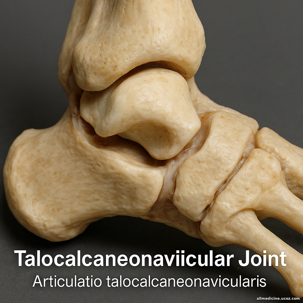
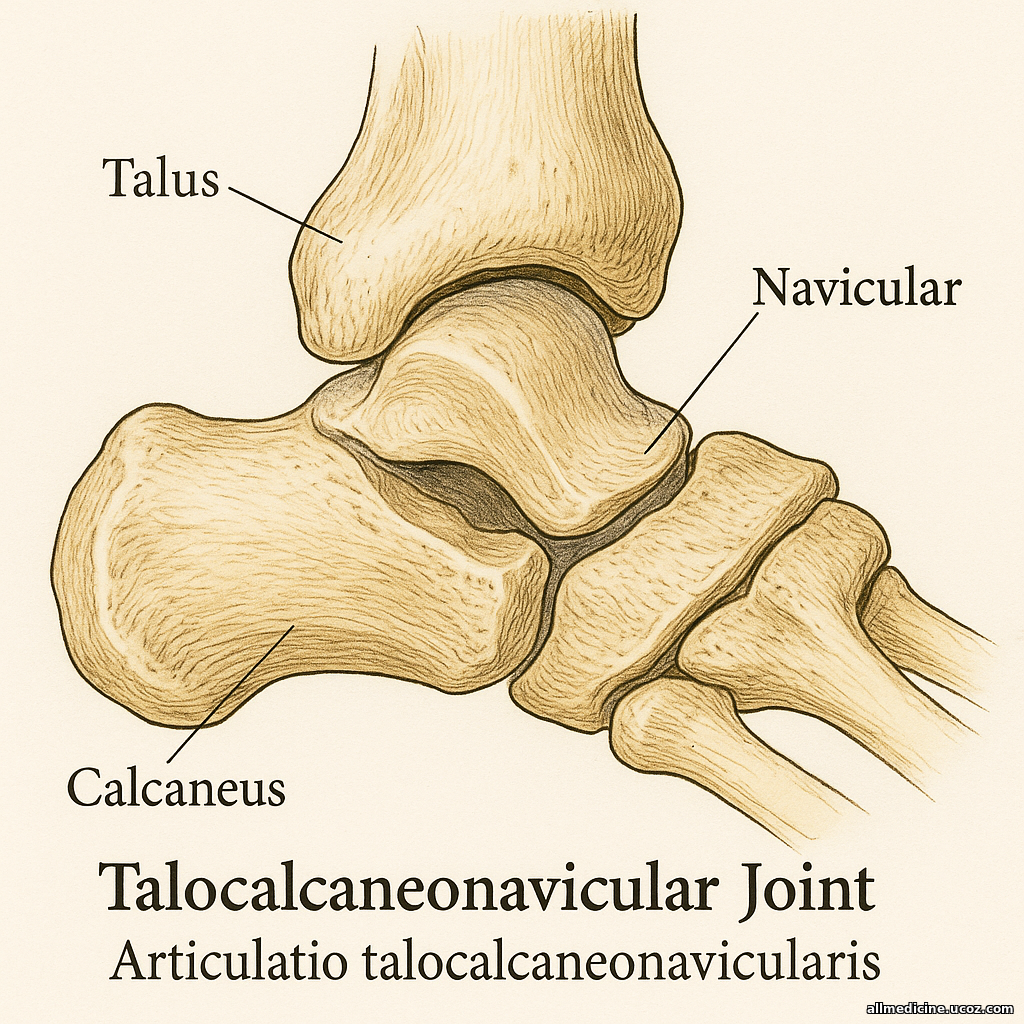
Talocalcaneonavicular Joint (Articulatio Talocalcaneonavicularis)
The talocalcaneonavicular joint is formed by the articular surfaces of the talus, calcaneus, and navicular bones. The head of the talus acts as the articular head, while the calcaneus and navicular bones together form the articular socket. The joint capsule (capsula articularis) attaches along the margins of the articular cartilage.
Ligaments reinforcing the joint:
-
Talonavicular ligament (lig. talonaviculare): Extends from the neck of the talus to the navicular bone.
-
Plantar calcaneonavicular ligament (lig. calcaneonaviculare plantare): Also known as the “spring ligament,” it stretches from the sustentaculum tali of the calcaneus to the plantar surface of the navicular bone. Its superior part blends with the navicular fibrocartilage, which contributes to the articular socket.
Although anatomically this is a spheroidal (ball-and-socket) joint, functionally it permits movement only in one plane, approximately in the sagittal direction, due to ligament constraints.
This joint is formed by the posterior articular surface of the cuboid bone (facies articularis posterior ossis cuboidei) and the cuboid articular surface of the calcaneus (facies articularis cuboidea calcanei). The articular surfaces are saddle-shaped.
The joint capsule is tightly attached along the cartilage edge on the medial side and looser on the lateral side, where it attaches slightly away from the cartilage.
Reinforcing ligaments (mainly plantar side):
-
Long plantar ligament (lig. plantare longum): The strongest ligament of this joint, arising from the plantar surface of the calcaneal tuberosity and running anteriorly. It arches over the groove of the cuboid (sulcus ossis cuboidei), forming a fibro-osseous canal, and inserts onto the bases of the 2nd to 5th metatarsal bones.
-
Deep fibers of this ligament insert onto the tuberosity of the cuboid bone.
-
-
Plantar calcaneocuboid ligament (lig. calcaneocuboideum plantare): Lies deeper, directly reinforcing the joint capsule, and connects the plantar surfaces of the calcaneus and cuboid.
Although anatomically saddle-shaped (articulatio sellaris), functionally this is a uniaxial joint.
Transverse Tarsal Joint (Articulatio Tarsi Transversa)
The transverse tarsal joint combines two joints:
-
Talocalcaneonavicular joint (articulatio talocalcaneonavicularis)
-
Calcaneocuboid joint (articulatio calcaneocuboidea)
The joint line forms an S-shaped curve:
-
Medial segment bulges anteriorly
-
Lateral segment curves posteriorly
Although anatomically separated, they are functionally linked by the bifurcate ligament (lig. bifurcatum).
Bifurcate ligament (lig. bifurcatum):
-
Originates from the dorsal surface of the anterior calcaneus and divides into two parts:
-
Calcaneocuboid ligament (lig. calcaneocuboideum): Passes to the dorsal surface of the cuboid.
-
Calcaneonavicular ligament (lig. calcaneonaviculare): Extends to the navicular bone.
-
This ligament acts as a “key” stabilizer of the transverse tarsal joint. Even if other surrounding ligaments are severed, the joint remains stable until this ligament is also cut, which is critical in surgical disarticulation of the foot at this level.
Tarsometatarsal Joints (Articulationes Tarsometatarseae)
These joints connect the tarsal bones to the metatarsal bones. There are three main articulations:
-
Between the medial cuneiform and the 1st metatarsal.
-
Between the intermediate and lateral cuneiforms and the 2nd and 3rd metatarsals.
-
Between the cuboid and the 4th and 5th metatarsals.
-
The joint between the medial cuneiform and 1st metatarsal has a weakly saddle-shaped articular surface.
-
The other joints have flat (plane) articular surfaces.
The joint line of the tarsometatarsal joints is irregular, since the 2nd metatarsal is longer than the others, and the lateral cuneiform projects slightly more anteriorly than the cuboid.
Joint capsule (capsula articularis) attaches to the cartilage margins and is reinforced by the following ligaments:
-
Dorsal tarsometatarsal ligaments (ligg. tarsometatarsea dorsalia): Located on the dorsal aspect.
-
Plantar tarsometatarsal ligaments (ligg. tarsometatarsea plantaria): Located on the plantar side.
-
Intermetatarsal ligaments (ligg. metatarsea interossea): Found between the bases of the metatarsals.
-
Interosseous cuneometatarsal ligaments (ligg. cuneometatarsea interossea): Connect the cuneiform bones to the metatarsals.
-
The medial interosseous ligament, connecting the medial cuneiform with the base of the 2nd metatarsal, acts as the “key” of the tarsometatarsal joints.
-
These joints are classified as amphiarthroses (limited mobility joints).